Offered by
Cytopathology Foundation Inc
About this shop
CF-LCI (Cytopathology Foundation- Low Cost Inititative)

CellBlockistry 101: CMAS #01
$17.99
*Price includes shipping (Paper back Book)
Monograph editor:
Vinod B. Shidham, MD, FRCPath, FIAC.
Cancer is a lethal disease with increasing numbers. Early diagnosis with minimally invasive modality is an important step in management. Cytopathology is commonly used for early diagnosis to follow-up at various stages during the treatment of cancer.
In addition to the study of cellular details in cytology preparations, the cytology specimens may have to be processed similar to the surgical biopsies for some special studies. Cell‑blocks of cytology specimens are comparable to the formalin‑fixed paraffin‑embedded (FFPE) tissue from surgical pathology specimens. They allow various elective ancillary studies on a variety of specimens through enhanced cytopathologic interpretation, including opportunity to perform immunohistochemistry, special stains, molecular tests, and other recently evolving studies such as proteomics.
CellBlockistry is the science studying chemistry and the art of achieving quantitatively and qualitatively improved cell‑blocks from different types of specimens. In addition to the role of CellBlockistry in clinical medicine in the field of cancer, it has a significant role in other non-cancer areas including infection and storage disorders such amyloidosis. it has an increasing role in veterinary sciences and various basic science research.
This pioneering book addresses the cell‑block making process and “cell‑blocking” related challenges with recent advances in improved interdisciplinary coordination for maximally optimal outcome with ongoing research in the field.
Potential readers:
Pathologists, cytopathologists, cytotechnologists, cytoprep technologists, and other fields associated with cell-blocks directly or indirectly including radiologists, laboratory personnel, histotechnologists, immunohistochemistry technologists, veterinary sciences, basic science researchers.

CYTOPATHOLOGIC DIAGNOSIS of SEROUS FLUIDS: CMAS #2
$51.99
*Price includes shipping (Paper back Book)
CYTOPATHOLOGIC DIAGNOSIS of SEROUS FLUIDS
Vinod Shidham (Editor)
Lester J Layfield (Co-editor)
The preface to the second edition by Dr. Ruth Katz is summarized here. This book provides an essential tool to aid in the diagnosis of serous effusions. It is lavishly illustrated. The chapters have been authored by experienced and expert pathologists of international repute with detailed cytologic descriptions. They are well organized with a comprehensive list of references. There are tables containing an algorithmic approach to the diagnosis of different entities throughout the book. It is both practical as well as extremely comprehensive and is strongly recommended for cytology professionals from beginning students to the most experienced practitioners. Overall this second edition, while quite similar to the first edition, has extra images with updated immunohistochemistry and molecular pathology.
The first twelve chapters are devoted to topics, ranging from benign reactive effusions, common metastatic carcinomas, and mesothelioma to the more unusual and rare non-epithelial neoplasms and hematolymphoid disorders. The last three chapters are more technical and provide guidelines for the use of flow cytometry to diagnose lymphoproliferative infiltrates in effusions, including tables describing major molecular genetic abnormalities found in lymphoma, acute leukemias, and soft tissue tumors.
Each chapter demonstrates classic examples of different neoplasms with their characteristic architectural and cellular features, easily relatable to the diagnostic cytologist. Examples of different entities are clearly displayed using both Giemsa and Papanicolaou stains. In addition, the use of cell-block tissue stained with H and E is commonly used. cell-blocks are essential to perform in order to refine the malignant diagnosis so that the precisely targeted therapy or biological therapy may be used.
The chapter dedicated to peritoneal washings cytology is an essential read with an excellent overview of the topic, including the pitfalls. The chapter covering hematolymphoid disorders is a comprehensive review of both benign and malignant lymphoid proliferations involving the serous cavities but also includes rare but important conditions. It also covers the differential diagnosis of non-neoplastic causes of effusions such as Tuberculosis. Cytomorphology and immunophenotype by both ICC and flow cytometry are covered with useful comments detailing differences from what one would expect to see in a tissue section or direct FNA smear.
Throughout the book and in different chapters there are examples of reactive mesothelial proliferations and adenocarcinomas that could also be confused with the cytologic appearance of certain subtypes of mesothelioma. A useful algorithm is provided that includes updated molecular pathology information to be used in making a definitive diagnosis of mesothelioma.
Another important chapter on appropriate specimen handling and optimal preparation. This chapter also details optimal specimen processing methods including direct smears, liquid-based cytology preparations, Cytospin, and filter preparations. Highly useful protocols for the different commonly used stains such as Romanowsky stains, and Diff-Quik stains are presented in tabular form. In addition, an extremely comprehensive catalog of cell-block techniques is provided including the use of Histogel, gelatin, agar, plasma-thrombin, collodion bag embedding, and Nano NextGen CelBloking™ kits. This chapter also outlines the 5 tier reporting system recently described for serous fluids.
The final chapter provides in-depth information regarding methods for ICC, and commonly used and less commonly used immunomarkers for diagnosing different malignancies, which are richly detailed regarding their derivation, which clones to use, and methods for antigen retrieval.
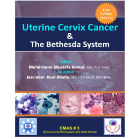
Uterine Cervix Cancer The Bethesda System: CMAS # 3
$42.99
*Price includes shipping (Paper back Book)
Uterine Cervix Cancer The Bethesda System: CMAS # 3
Meherbano Mustafa Kamal (Editor)
Jasvinder Kaur Bhatia (Co-Editor)
Vinod Shidham (Series Editor)
“Cancer Uterine Cervix & The Bethesda System” is a multi author book which discusses Cervical Cancer in its entirety. When the work of this book began almost seven
years back, the plan was to put together a practical compact format of a handbook of cervical cytology which would be useful in the diagnostic work at the microscope.
The aim was to focus mainly on the description of lesions, along with carefully selected illustrations, that a cytopathologist was likely to need in routine practice. As the contents progressed, there was an irresistible incentive to cover along with cytology, descriptions of histopathologic findings too. Having quick and easy reference material of histopathologic basis of cytology would be a very logical content of any handbook. This inevitably led to an expansion of text and illustrations to include method of collection of
Pap smear and screening technologies for cervical cancer with helpful feature of algorithms clearly intended to lead the way to standard follow up of women screened. Basics of Colposcopic findings in precancerous lesions of the cervix, that have bearing in the completion of the knowledge and workup of patients, have also been incorporated. As a result, the hand book has taken on the presentation of a textbook while still retaining the configuration of an Atlas because of its innumerable illustrations that have resulted in worthwhile increased coverage and usefulness New trends in the practice of gynecologic cytology have emerged. Therefore, a chapter of ancillary techniques, in particular immunocytochemistry using the
LBC smears was added. We have tried to satisfy the needs of the cytotechnicians within the set framework and format of any handbook by adding the chapters on
laboratory aspects that describe automation of cervical cytology and method of Papanicolaou staining and its nuances. This will ensure a wider range of receivers of
this book like the cytotechnologists and pathologists in general. For this reason alone, it deserves a place in the long history of development of meaningful cytopathologic literature in the field of cervical cancer.
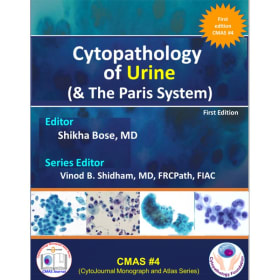
Cytopathology of Urine (& The Paris System): CMAS # 4
$64.99
*Actual reduced book price is $34.99.
Price includes international shipping and handling (Paper back Book)
For address witin Bharat, shipping cost of $30 is waived (please apply Code BHARAT$4- please dot apply this code for other CMAS books).
This monograph on urine cytology was designed with the aim of providing a comprehensive resource for pathologists, cytotechnologists, cytopathology trainees, and all those associated with managing urothelial pathologies. Cytopathology Monograph Atlas Series (CMAS) books are platform for availability of scientific cytopathology knowledge in open access on various cytopathology topics.
Add a donation for Cytopathology Foundation Inc
$
Did you know? We fundraise with Zeffy to ensure 100% of your purchase goes to our mission!